Elbow dysplasia gets far less attention than hip dysplasia, but for Labradors it’s arguably just as important to know about — the two conditions are frequently confused, yet they involve different joints, different diagnostic approaches, and different surgical options. This guide covers what elbow dysplasia actually is, how to recognize it early, and the full range of treatment paths available today.
- Defining Elbow Dysplasia and Its Four Subtypes
- Why Labradors Are Genetically at Risk
- Signs and Symptoms
- Diagnosis
- Treatment Options
- Managing Secondary Arthritis Long-Term
- Prognosis and Lifestyle Adjustments
- Prevention, Screening, and Breeding Considerations
- Elbow vs. Hip Dysplasia: Quick Comparison
- Frequently Asked Questions
Defining Elbow Dysplasia and Its Four Subtypes (FCP, UAP, OCD, Incongruity)
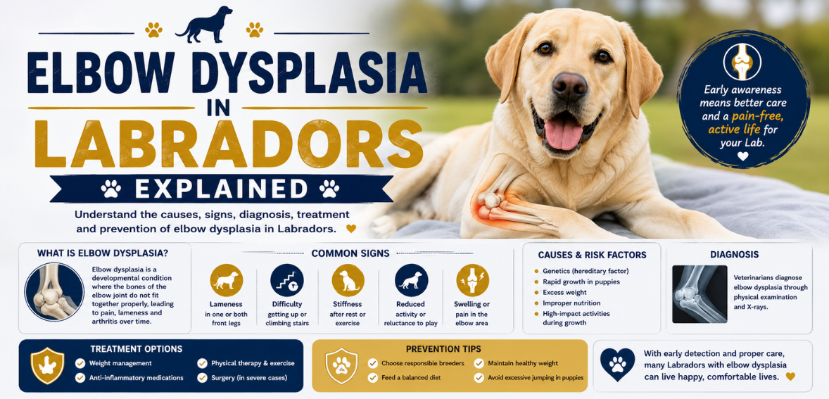
“Elbow dysplasia” is an umbrella term covering four distinct developmental problems in the elbow joint, which is formed where the humerus, radius, and ulna meet. Any one — or a combination — can occur in the same dog, and up to 80% of affected dogs have the condition in both elbows.
| Subtype | What’s Happening | Typical Treatment Approach |
|---|---|---|
| Fragmented Coronoid Process (FCP) | A small bony process on the ulna cracks and separates — the most common subtype | Arthroscopic fragment removal |
| Ununited Anconeal Process (UAP) | A bony projection on the ulna fails to fuse to the rest of the bone at puberty | Open surgery (arthrotomy) — fragment removal or screw reattachment |
| Osteochondrosis (OCD) of the medial humeral condyle | Cartilage fails to properly convert to bone, leaving a thicker, unstable cartilage layer that can flake off | Arthroscopic removal of loose cartilage |
| Elbow incongruity | The bones of the joint don’t fit together with normal alignment, creating abnormal pressure points | May require an ulna osteotomy to correct joint alignment |
Why Labradors Are Genetically at Risk
According to the American College of Veterinary Surgeons (ACVS), Labrador Retrievers are among the breeds with the highest predilection for “medial compartment disease” (the FCP/OCD/incongruity group), alongside Golden Retrievers and German Shepherds. The exact cause is believed to be multifactorial — a genetic predisposition combined with rapid growth rate, body weight, and possibly diet during the puppy growth period, though genetics is considered the dominant factor. This is exactly why OFA elbow screening of breeding stock matters for reducing the condition’s prevalence in the breed over time.
Signs and Symptoms (Front-Leg Lameness, Worse After Rest/Exercise)
- Front-leg (forelimb) lameness, sometimes affecting both front legs, usually first noticed between 4 and 8 months old
- Stiffness after rest that may improve slightly with gentle movement, then worsen again after strenuous exercise
- Reluctance to fully extend or flex the elbow, sometimes visible as a shortened stride in the front legs
- Elbows held slightly out from the body rather than tucked close, in some affected dogs
- Owners often initially mistake the lameness for a simple sprain, since it can wax and wane rather than being constantly obvious
Any front-leg limping lasting more than a day or two, especially in a puppy between 4 and 8 months old, or lameness that consistently worsens after exercise. Early diagnosis meaningfully widens treatment options and improves long-term outcomes.
Diagnosis (X-ray, CT, Arthroscopy, OFA Elbow Grades)
- X-ray: The OFA requires a specific extreme flexed mediolateral view, looking especially for new bone growth (osteophytes) on the anconeal process — often the most sensitive early radiographic sign
- CT scan: Provides more detailed cross-sectional imaging than standard X-rays, often used when FCP or subtle incongruity is suspected but not clearly visible on X-ray
- Arthroscopy: A minimally invasive procedure using a small camera inside the joint — often considered the most accurate way to diagnose FCP and OCD specifically, and can treat the problem in the same procedure
- OFA elbow grading: Dogs are graded Normal, or Grade I, II, or III (mild, moderate, or severe) based on the degree of arthritic change visible on X-ray
Treatment Options (Arthroscopic Surgery, Medical Management)
Treatment depends on which subtype is present, the dog’s age, and how much secondary arthritis has already developed.
- FCP and OCD: Usually treated arthroscopically, removing the loose fragment or unstable cartilage
- UAP: Typically requires an open surgical approach (arthrotomy) to remove or screw-reattach the ununited fragment
- Elbow incongruity: May require an ulna osteotomy to re-establish normal joint alignment
- Medical management (weight control, NSAIDs, physical therapy, joint supplements) may be recommended instead of surgery for very mild cases, or when arthritis has already progressed to the point surgery offers limited additional benefit
Surgical cost typically runs $1,500 to $5,000 per elbow, varying by region, specialist, and procedure complexity; since the condition affects both elbows in a majority of cases, budgeting for bilateral treatment is often necessary. Elbow arthroscopy is generally performed by a board-certified veterinary surgeon (ACVS diplomate) rather than a general practice vet.
Managing Secondary Arthritis Long-Term
Whether or not surgery is performed, elbow dysplasia commonly leads to some degree of osteoarthritis over time, since the initial joint damage changes how the joint bears weight for the rest of the dog’s life. Long-term management typically includes maintaining a lean body weight, controlled low-impact exercise like swimming, joint supplements, and NSAIDs or other pain management during flare-ups, all under ongoing veterinary guidance.
Prognosis and Lifestyle Adjustments
Outcomes vary by subtype, severity, and how early treatment begins. Published outcomes for arthroscopic surgery show roughly 40% of dogs return to essentially normal function, another 30–35% achieve a good-to-fair outcome with some ongoing management needed, and a smaller portion (5–10%) show limited improvement — often those with more advanced arthritis already present at the time of surgery. This underscores why early diagnosis matters so much: dogs treated before significant secondary arthritis develops generally do better than those diagnosed later.
See our guide to exercise and joint health for Labs for how to adjust activity for a dog managing this condition long-term.
Prevention, Screening, and Breeding Considerations
- OFA elbow evaluation of both parents before breeding is the primary prevention tool, since genetics is the leading risk factor
- Ask breeders for verifiable OFA elbow grades, checkable on the OFA public database, not just verbal assurance
- Moderate, steady puppy growth via a large-breed puppy formula with controlled calcium and phosphorus, rather than rapid growth from overfeeding
- Maintain lean body condition throughout the dog’s life to reduce joint stress
See our guide to Labrador puppy growth and nutrition for how feeding choices support healthy joint development, and our full guide to common Labrador health problems for the broader picture of breed-specific health.
Elbow vs. Hip Dysplasia: Quick Comparison
| Elbow Dysplasia | Hip Dysplasia | |
|---|---|---|
| Joint affected | Front leg (elbow) | Back leg (hip) |
| Typical age of first signs | 4–8 months | 5 months–2 years, sometimes later |
| Classic gait sign | Front-leg lameness, shortened stride | “Bunny hopping,” rear-end sway |
| OFA screening | Elbow-specific extreme flexed view | Hip-specific standard view or PennHIP |
See our full guide to hip dysplasia in Labradors for the equivalent breakdown of that condition.
Frequently Asked Questions
What is elbow dysplasia in Labradors?
A group of developmental elbow joint conditions — FCP, UAP, OCD, and joint incongruity — where the bones don’t form or fit together properly during growth, causing front-leg lameness and, over time, arthritis. Labradors are among the breeds most predisposed.
How do I know if my Lab has elbow dysplasia?
Watch for front-leg lameness, especially in a puppy between 4 and 8 months old, stiffness after rest that changes with exercise, and a shortened stride. A vet visit with X-rays is needed to confirm diagnosis.
Is elbow dysplasia in dogs curable?
Not always fully “curable,” but it’s very manageable. Surgery can significantly improve function, especially when performed early, though some degree of secondary arthritis often develops over time regardless of treatment.
What’s the difference between hip and elbow dysplasia?
Elbow dysplasia affects the front legs and typically shows up earlier (4-8 months), causing front-limb lameness, while hip dysplasia affects the back legs and hips, often with a “bunny hopping” gait, and can show up anywhere from 5 months to later in life.
How much does elbow dysplasia surgery cost?
Typically $1,500 to $5,000 per elbow, depending on the specific procedure, region, and specialist. Since both elbows are affected in the majority of cases, many owners budget for bilateral treatment.
Want to understand how this compares to the other major joint condition in the breed?
Read our full guide to hip dysplasia in Labradors →Sources
- American College of Veterinary Surgeons (ACVS) — Canine Elbow Dysplasia
- Orthopedic Foundation for Animals (OFA) — Elbow Dysplasia Grading Standards
- American Kennel Club — Elbow Dysplasia in Dogs: Signs, Symptoms, Treatment
- PetMD — Elbow Dysplasia in Dogs: Symptoms, Causes, and Treatment